Challenge (Level 5)

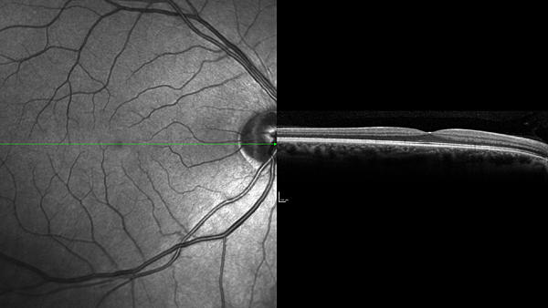
An infrared scan and cross-section of Mark's right retina!
These are images of my retina (the back surface of my right eye). In the left panel you can see the blood vessels, the beginning of the optic nerve (right edge of image) and my fovea (near the center with no blood vessels). The right panel is a cross section of the area marked with the green line. You can see a slice through my fovea (the pit where the nerve fibers are pulled away to allow better vision) and the various layers of cells in my retina. The rod and cone inner segments are the dark gray area near the middle of the cross section.


Quiz Time: What Is the Functional Difference Between Rods and Cones?


(A) Nothing; They Are Exactly the Same

(B) Cones Hold More Ice Cream

(C) Cones Are Capable of Detecting Color; Rods Are Not

(D) Cones Respond to Light; Rods Respond to Dark


FEEDBACK

Updated: July 29, 2010